

Services
The Fluorescence Microscopy and Imaging Center provides intramural researchers with access to a variety of advanced imaging equipment and techniques that will further their research. The center’s experienced staff also offers training on image acquisition and data analysis.
Background
What is fluorescence microscopy?
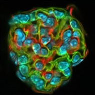
Fluorescence microscopy is a non-destructive way of tracking or analyzing tissues, cells, or cellular structures. The procedure includes labeling an antibody with a fluorescent dye known as a fluorophore, and allowing it to find its target antigen within a sample. Fluorophores may also be covalently bonded to small molecules or proteins. Samples are visualized using an array of microscopic methods.
Types of equipment we use:
- Zeiss LSM 980 confocal microscope with Airy-scan
- Zeiss LSM 880 confocal microscope with Airy-scan
- Zeiss LSM 780 confocal microscope
- Zeiss LSM 710 confocal and multiphoton microscope
- Zeiss Lightsheet 7
- Andor Dragonfly (spinning disk confocal, TIRF, dSTORM)
- Olympus and Zeiss epifluorescence microscopes
- Image Analysis workstations
NIEHS Shared and Core Facilities are available to NIH researchers. Information for staff on utilizing these services may be found on the NIEHS Junction or by contacting the staff below.
Video
Scientific Staff
-

-
Charles J. Tucker
Director, Fluorescence Microscopy and Imaging Center -
P.O. Box 12233Mail Drop F2-09Durham, NC 27709
-
Tel 984-287-3469
[email protected]
-

-
Erica L. Scappini, M.S.
Biologist -
P.O. Box 12233Mail Drop F2-09Durham, NC 27709
-
Tel 984-287-3468
[email protected]
-

-
Robert N. Wine, M.S.
Biologist -
P.O. Box 12233Mail Drop F2-09Durham, NC 27709
-
Tel 984-287-3551
[email protected]