

The Digitized Atlas of Mouse Liver Lesions
![]()
Much of the work carried out by DTT is in support of the National Toxicology Program (NTP), an interagency partnership of the Food and Drug Administration, National Institute for Occupational Safety and Health, and NIEHS.
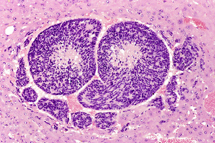
Hepatoblastomas may be small or large lesions comprised of hyperchromatic cells with scant cytoplasm. Most appear to arise within hepatocellular carcinomas and occasionally within adenomas as well demarcated lesions. It is speculated that hepatoblastomas represent a poorly differentiated variant of hepatocellular carcinomas.
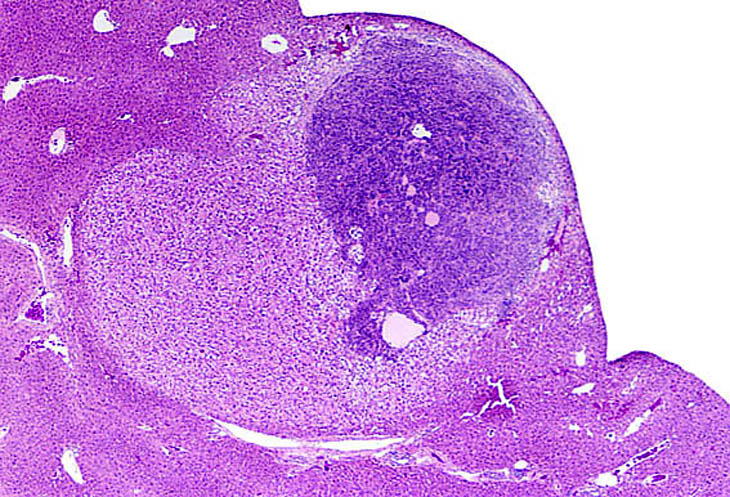
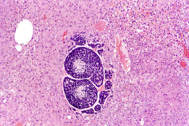
A basophilic hepatoblastoma is arising within a discrete hepatocellular adenoma.
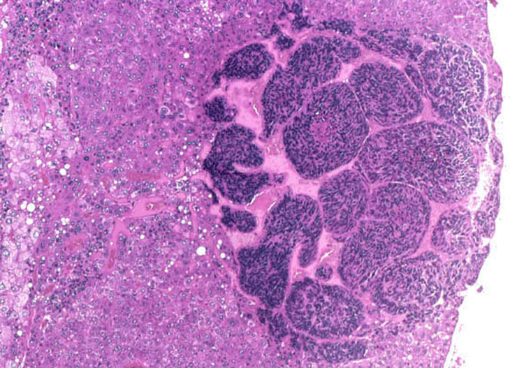
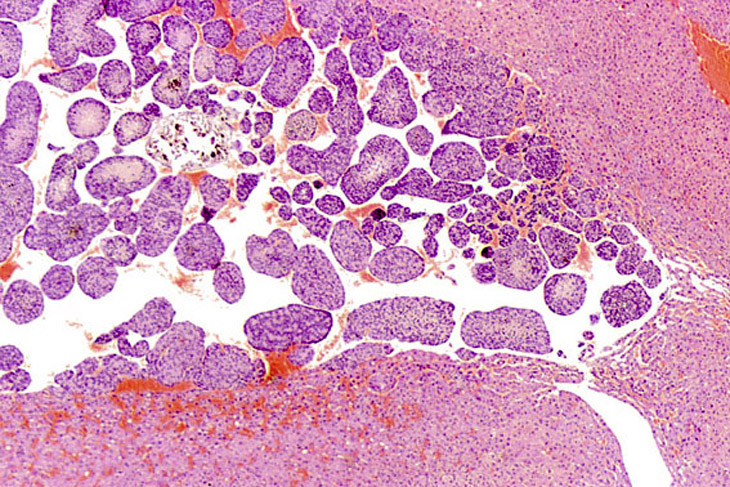
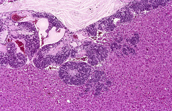
This small hepatoblastoma is present within an hepatocellular carcinoma.
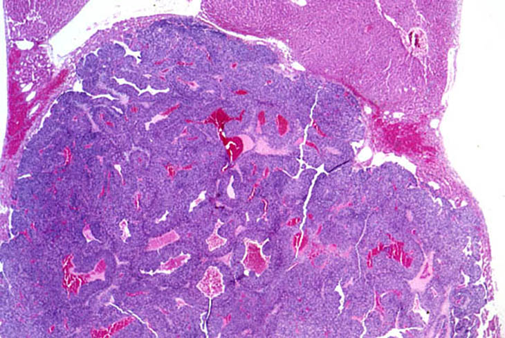
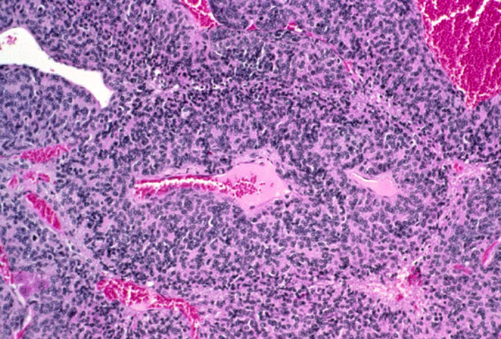
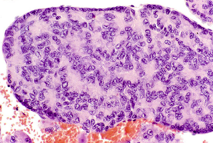
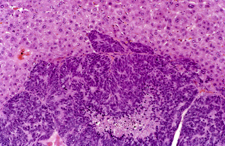
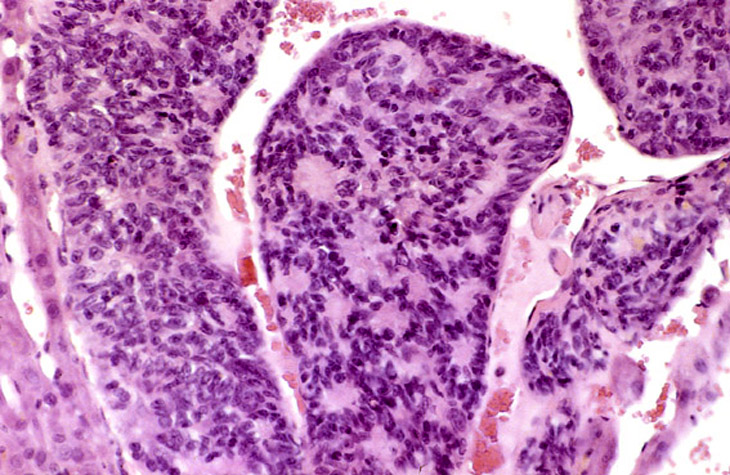
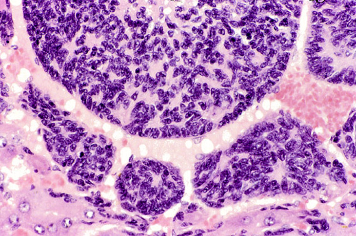
A large hepatoblastoma; higher magnification shows malignant cells palisading around vascular structures.
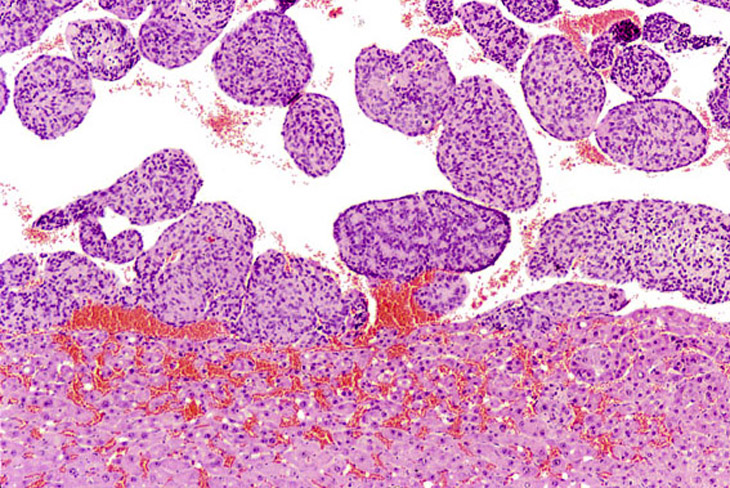
Hepatoblastoma associated with hemorrhage.
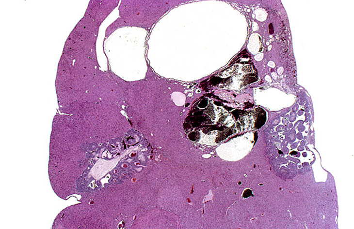
Hepatoblastomas and biliary cysts in the liver of an adult mouse.
A small hepatoblastoma within an hepatocellular carcinoma. Non-neoplastic hepatic parenchyma is present in the upper right.