The Digitized Atlas of Mouse Liver Lesions
Much of the work carried out by DTT is in support of the National Toxicology Program (NTP), an interagency partnership of the Food and Drug Administration, National Institute for Occupational Safety and Health, and NIEHS.
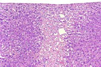
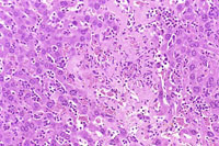
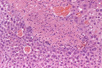
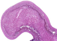
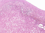
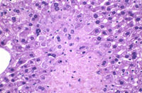
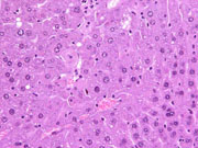
Coagulation necrosis is characterized by distinct eosinophilic cytoplasm with pyknotic or absent nuclei. Infarction of hepatic lobes is a form of coagulation necrosis.
Focal coagulation necrosis.
Another example of focal necrosis.
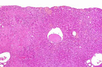
Extensive necrosis of a liver lobe may occur secondary to torsion.
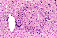
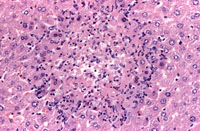
The left image shows a focus of necrosis with an early inflammation cell infiltration at the periphery of the necrotic hepatocytes. In the right image, the inflammatory reaction is more pronounced.
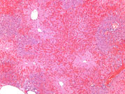
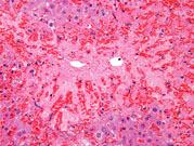
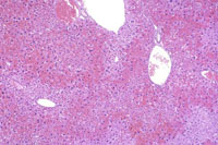
This is an example of centrilobular hepatocellular necrosis with associated hemorrhage sometimes referred to as hemorrhagic necrosis.
Marked centrilobular necrosis in a B6C3F1 male mouse chronically fed acetaminophen. Note depression of liver surface due to hepatocyte loss and pigment deposition. Areas of hemorrhage are often associated with the necrosis.
In addition to hepatic necrosis, inflammation, and pigment deposition, there is a venous thrombus present in this mouse treated with acetaminophen.
Necrosis and hemorrhage are present in this mouse treated with acetaminophen.
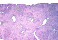
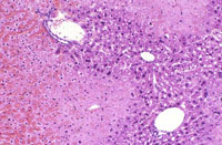
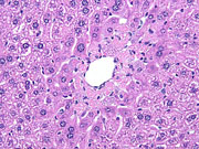
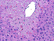
Mild centrilobular necrosis is evident following treatment with acetaminophen.
Another example of mild centrilobular necrosis induced by acetaminophen treatment. Note nuclear and cytoplasmic changes in hepatocytes surrounding the central vein.
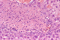
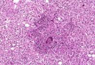
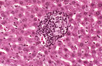
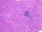
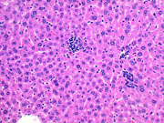
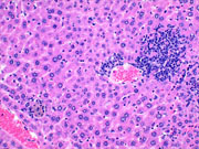
Microgranulomas consisting of mononuclear inflammatory cells and associated with hepatocyte necrosis are commonly seen in mouse liver.
Granuloma in the liver of a B6C3F1 mouse.
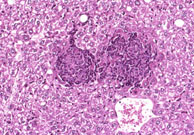
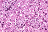
Hepatic granulomas in a mouse given dietary tetrachlorvinphos for 2 years. Note that these granulomas are comprised primarily of macrophages and multinucleated giant cells.
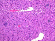
Multiple granulomas are present in this Tg.AC mouse treated with rotenone.


