

The Digitized Atlas of Mouse Liver Lesions
![]()
Much of the work carried out by DTT is in support of the National Toxicology Program (NTP), an interagency partnership of the Food and Drug Administration, National Institute for Occupational Safety and Health, and NIEHS.
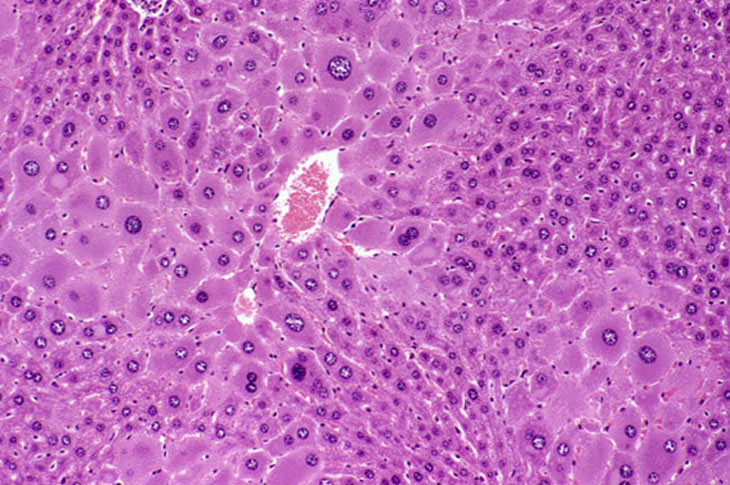
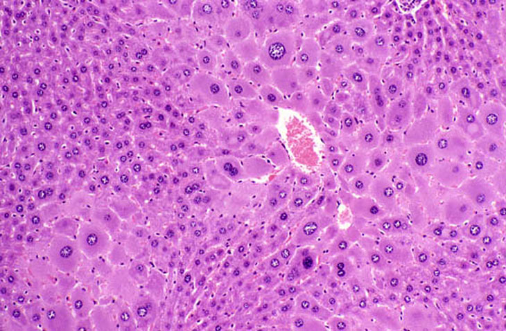
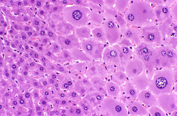
Hepatocellular hypertrophy is frequently seen in the liver following exposure to agents that cause hepatic enzyme induction. The hypertrophy typically starts in the centrilobular area and extends to the mid-lobular and eventually periportal areas provided there is sufficient stimulus over time. In situations of prolonged exposure to some agents, the hypertrophic hepatocytes are seen to have enlarged polyploid nuclei. With some treatment regimens, hepatocytes may actually become cytomegalic. Cytomegalic hepatocytes typically have single polyploid and/or multiple nuclei.
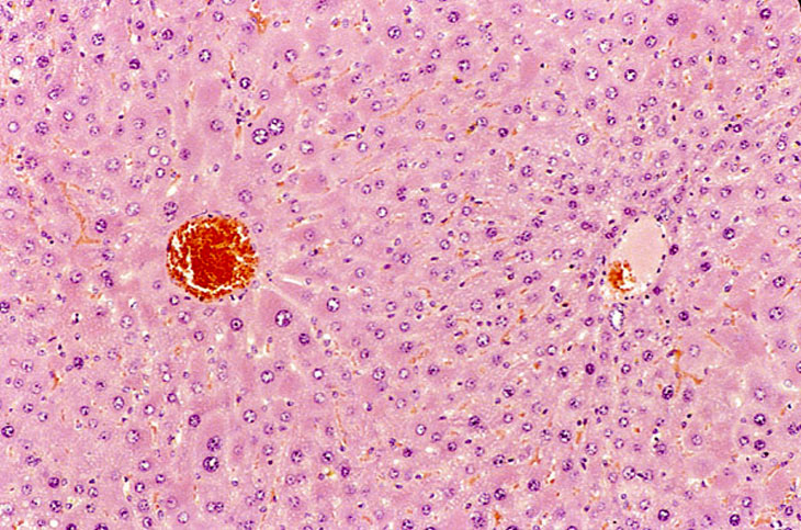
Minimal centrilobular hepatocyte hypertrophy is present and most dramatically involves enlargement of hepatocytes immediately adjacent to the central vein. The periportal hepatocytes (right side of image) are not affected.
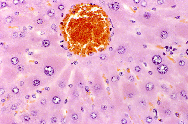
Generalized hepatocyte hypertrophy involving all portions of the hepatic lobule. Higher magnification shows a cytomegalic hepatocyte in the lower right. It has an enlarged nucleus with multiple nucleoli.
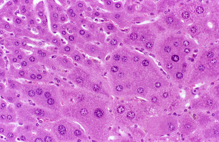
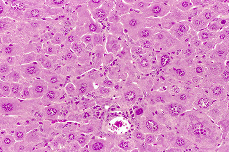
Hypertrophic and cytomegalic hepatocytes are present in the centrilobular and midlobular areas in these images from a mouse treated with chlordane for several months. Some cytomegalic hepatocytes have several nuclei.