Services
The Flow Cytometry Center provides state of the art instrumentation and resources for fluorescent analysis of cells at the single cell level, including high-speed fluorescence-activated cell sorting. Our goal is to provide expertise in flow cytometry in the areas of experimental design, instrument knowledge and training, data analysis and cell sorting. We support the mission of NIEHS by providing the latest advances in flow cytometric technology to the institute for reducing the burden of diseases and dysfunctions associated with the environment.
Background
Cells stained with various fluorescent markers or dyes pass single file by a set of lasers where the cells of interest are specifically selected and sorted or isolated into individual tubes for further biochemical analysis. (NOTE: Animation is video only and has no audio.)
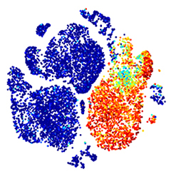
Flow cytometry is a powerful technique to simultaneously analyze multiple characteristics of thousands of individual cells in a relatively short period of time. Unlike other biochemical techniques, flow cytometry makes these multiparametric measurements on single cells as opposed to population measurements. Cells stained with various fluorescent markers or dyes pass single file by a set of lasers where the cells of interest are analyzed and/or specifically selected and sorted (isolated) into individual tubes for further biochemical analysis.
Recent technical advances blended elements of flow cytometry and time-of-flight mass spectrometry into what has become known as mass cytometry. Unlike flow cytometry which has parametric limitations due to fluorescence spill-over, mass cytometry’s use of metal-labeled antibody probes to measure dozens of parameters at a single cell level. With 135 detection channels, the Helios mass cytometer can simultaneously resolve multiple elemental probes at high acquisition rates, thereby maximizing the per-cell information obtained from a single sample. The Hyperion upgrade allows for this type of analysis directly on tissue samples permitting special orientation of the markers of interest.
Instrumentation:
- BD LSRFortessa
- BD FACSAriaII
- BD Symphony S6
- BD FACSMelody
- Sony ec800
- Helios/Hyperion mass cytometry system
NIEHS Shared and Core Facilities are available to NIH researchers. Information for staff on utilizing these services may be found on the NIEHS Junction or by contacting the staff below.
Video
Scientific Staff
Carl D. Bortner, Ph.D.