


The Digitized Atlas of Mouse Liver Lesions
Much of the work carried out by DTT is in support of the National Toxicology Program (NTP), an interagency partnership of the Food and Drug Administration, National Institute for Occupational Safety and Health, and NIEHS.
Visit the NTP WebsiteThe presence of erythrocytes within hepatocytes is seen on rare occasions. It is not known for sure why or how intact erythrocytes come to be located within individual hepatocytes. Perhaps it is by erythrophagocytosis, but that is only supposition. The affected hepatocytes often become very large and, presumably, ultimately die and become phagocytosized by Kupffer cells along with their contents. Exacerbation of this change by chronic treatment may occur. We have seen it in three of approximately 500 NTP two-year studies. The cause and significance of this change is not known.
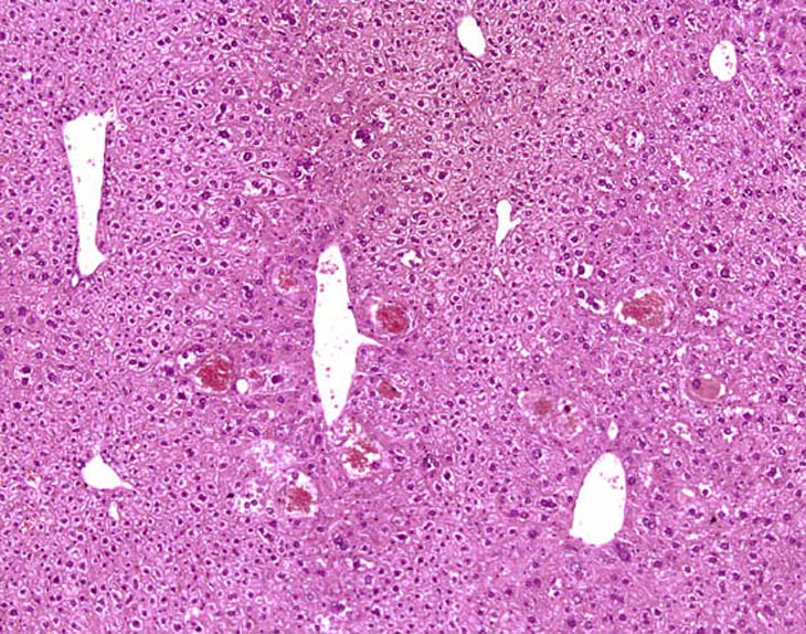
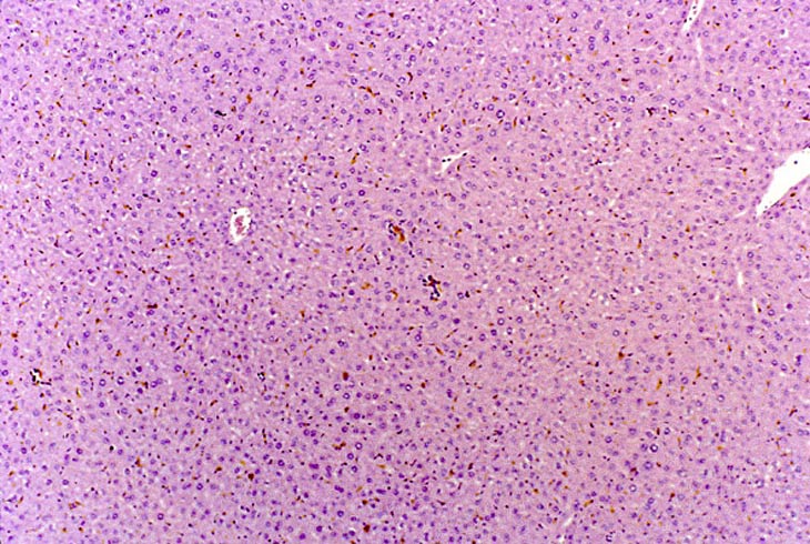
Low magnification of a liver in which multiple small collections of erythrocytes that resemble peliosis can be seen. These actually represent individual enlarged hepatocytes containing numerous erythrocytes.

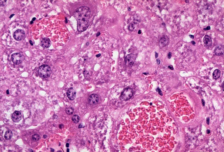
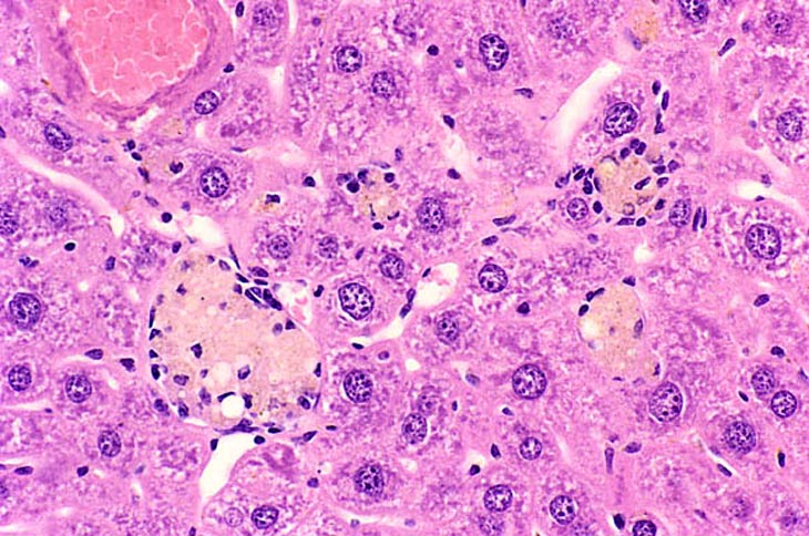
Several hepatocytes contain erythrocytes. The nucleus is of normal size while the cytoplasmic mass is markedly enlarged with margination of the cytoplasm. The nucleus in these cells is often localized at the cell margin.
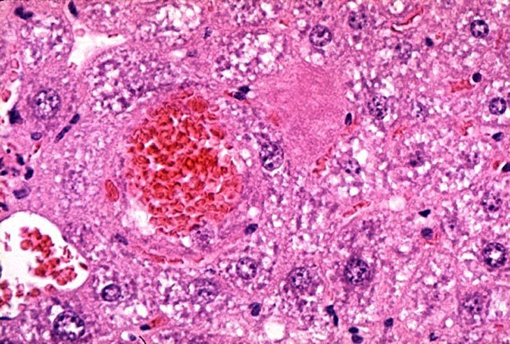
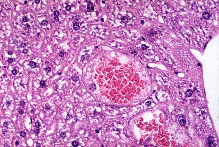
Intrahepatocytic erythrocytes.
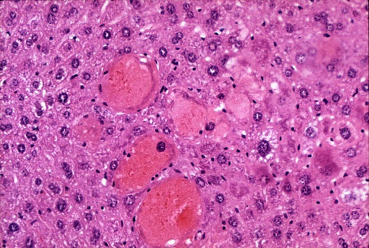
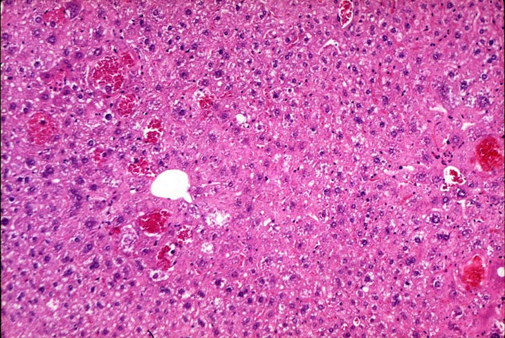
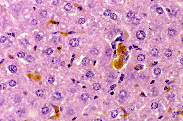
In this example, the intrahepatocytic erythrocytes have become partially lysed.
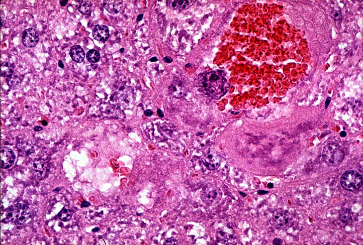
In addition to an hepatocytes filled with erythrocytes, there is accumulation of pigment within Kupffer cells (macrophages).
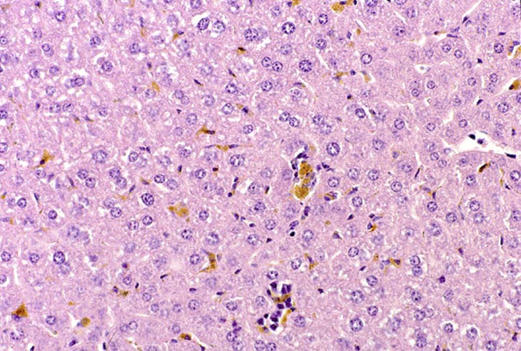
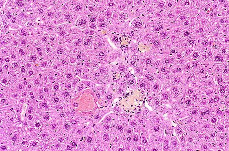
Kupffer cells throughout the liver are filled with golden brown pigment which is believed to represent breakdown of hemoglobin.