


The Digitized Atlas of Mouse Liver Lesions
Much of the work carried out by DTT is in support of the National Toxicology Program (NTP), an interagency partnership of the Food and Drug Administration, National Institute for Occupational Safety and Health, and NIEHS.
Visit the NTP Website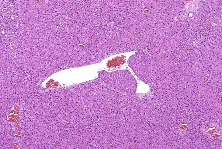
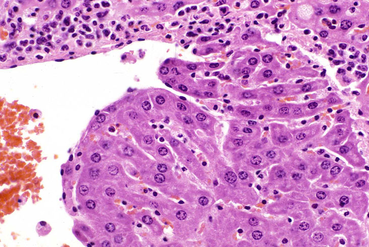
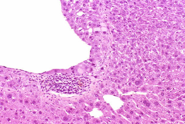
In some foci of cellular alteration, primarily basophilic foci, hepatocytes may occasional be seen to protrude into the lumen of hepatic veins. They are usually lined by a layer of flattened endothelial cells. This change has been considered by some to represent a form of microinvasion and such lesions have been diagnosed as hepatocellular carcinomas, possibly motivated by the fact that most are seen within foci induced by treatment with hepatocarcinogens. However, similar changes are occasionally seen in untreated mice and may not necessarily be associated with a focus of cellular alteration. Consequently, we cannot be certain that this change is actually a microcarcinoma. Two clusters of hepatocytes protruding into a large hepatic vein.
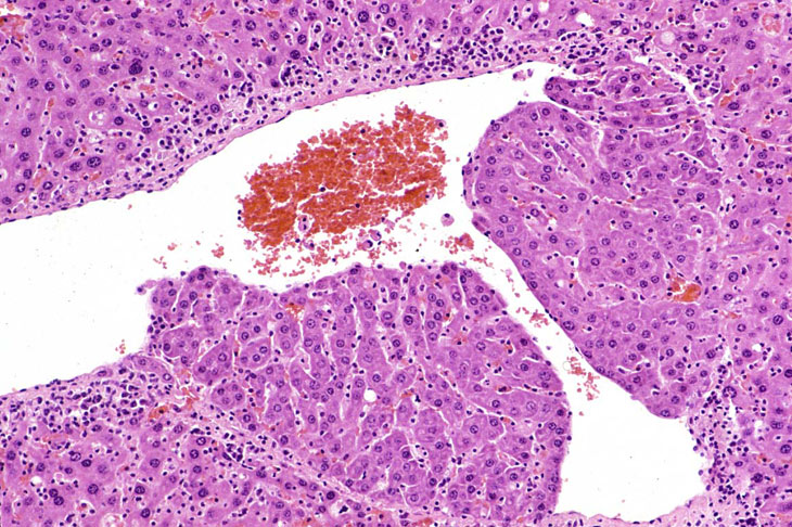
Another example of protrusion of hepatocytes into an hepatic vein.