


The Digitized Atlas of Mouse Liver Lesions
Much of the work carried out by DTT is in support of the National Toxicology Program (NTP), an interagency partnership of the Food and Drug Administration, National Institute for Occupational Safety and Health, and NIEHS.
Visit the NTP Website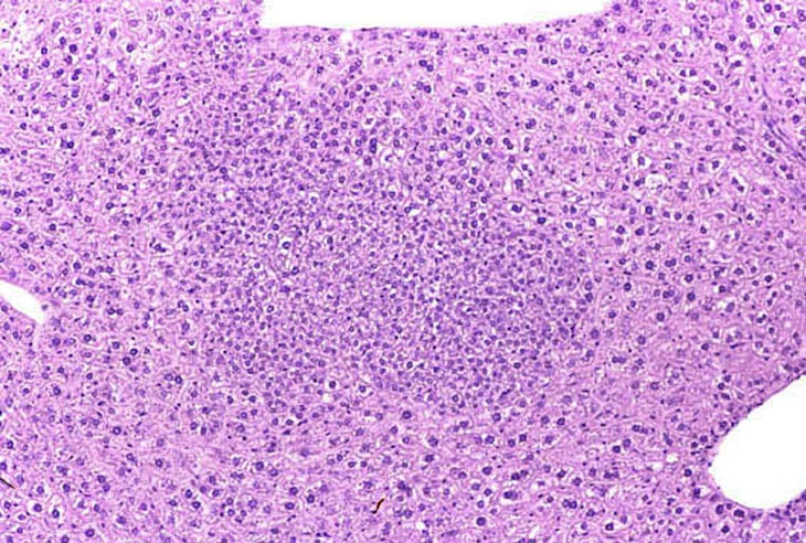
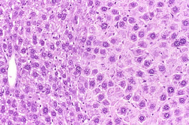
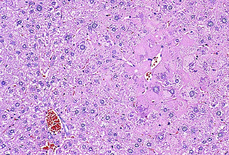
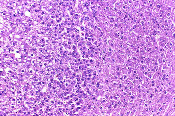
Foci of cellular alteration represent small to large aggregates of tinctorially distinct hepatocytes within the hepatic parenchyma and are sometimes considered putative preneoplastic lesions. They are frequently classified based upon their phenotypic appearance as basophilic, eosinophilic, clear cell, vacuolated, and mixed foci. Occasionally amphophilic foci are identified by the altered arrangement of the hepatic plates without tinctorial distinction from the surrounding hepatic parenchyma. The distinction between a large focus of cellular alteration and a hepatocellular adenoma is often a difficult judgment. A basophilic focus of cellular alteration with irregular boundaries. Basophilic foci are frequently comprised of hepatocytes that are smaller than the surrounding hepatocytes. The tinctorial properties are a function of closely spaced small nuclei and/or increased cytoplasmic basophilia.

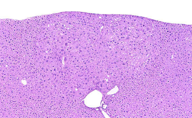
Multiple basophilic foci are prominent in this mouse treated with a neonatal dose of diethylnitrosamine.
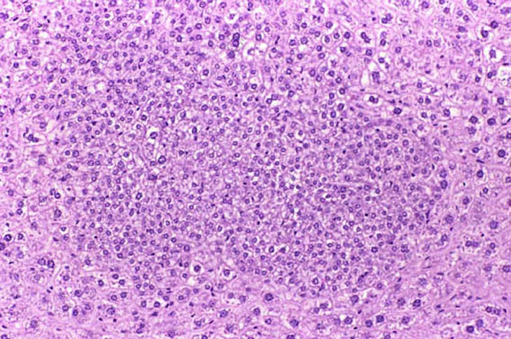
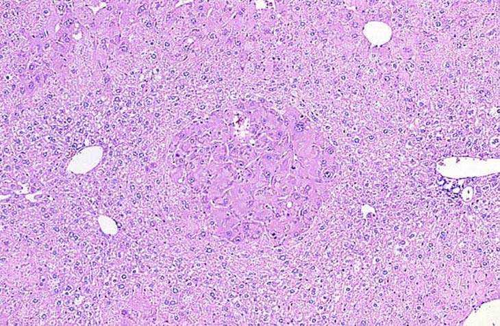
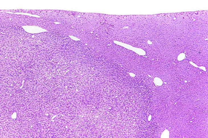
A large eosinophilic focus of cellular alteration that has a well demarcated border. Hepatocytes comprising eosinophilic foci typically have an increased cytoplasm that stains more eosinophilic than the cytoplasm of surrounding hepatocytes. If the hepatocytes within an eosinophilic focus are sufficiently large and numerous, there may be evidence of slight compression of normal hepatic parenchyma along a portion of the border of the focus.
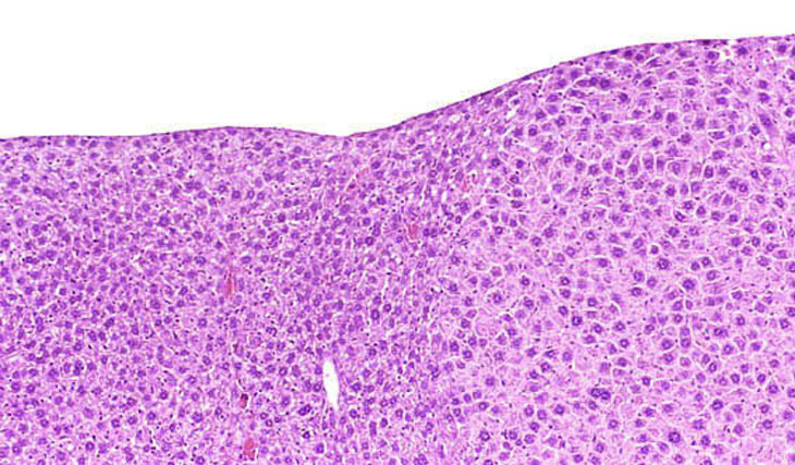
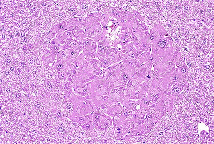
An eosinophilic focus of cellular alteration with slight protrusion above the normal surface contour of the liver.
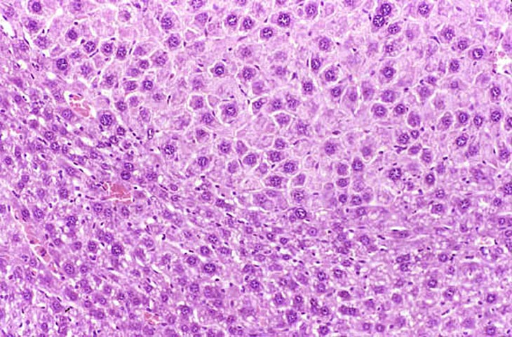
A well delineated small eosinophilic focus comprised of hepatocytes with copious cytoplasm.
A small eosinophilic focus of cellular alteration with an irregular border.
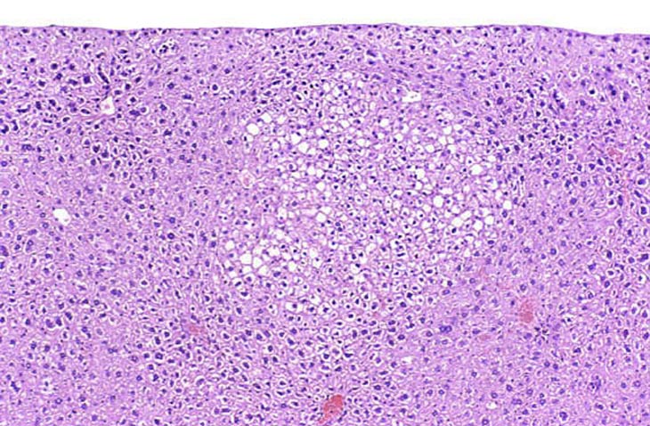
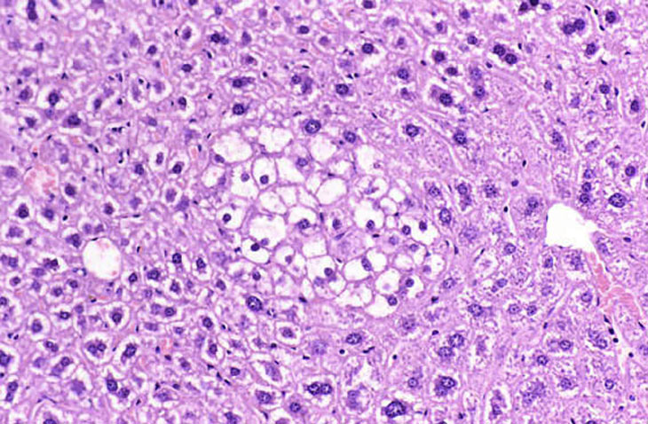
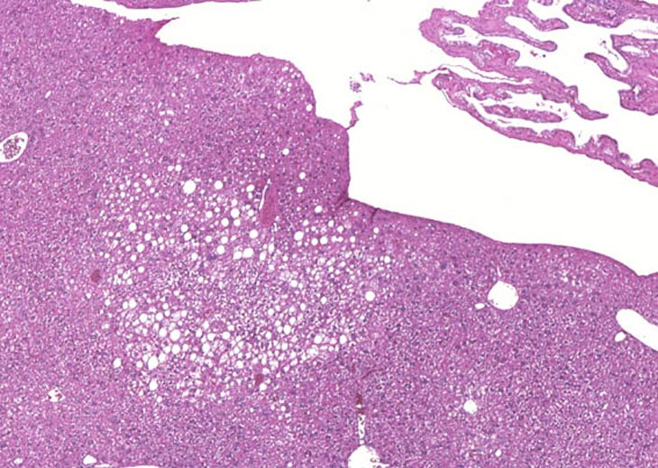
A large and a small clear cell focus of cellular alteration. Clear cell foci are characterized by relatively clear cytoplasm or cytoplasm with just a hint of very pale eosinophilic staining and wispy strands of cytoplasm making the cytoplasmic vacuoles have an indistinct border. Unlike vacuolated foci, many cells within a clear cell focus have a centrally located nucleus. The clear space is produced when stored glycogen is dissolved out during fixation in aqueous fixatives.
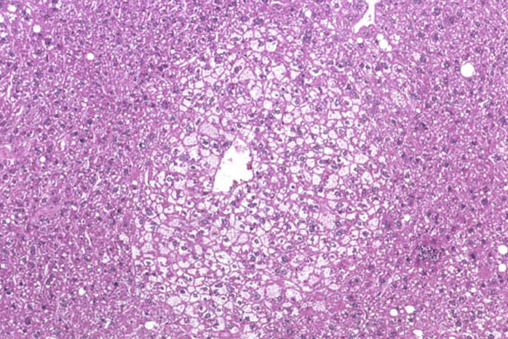
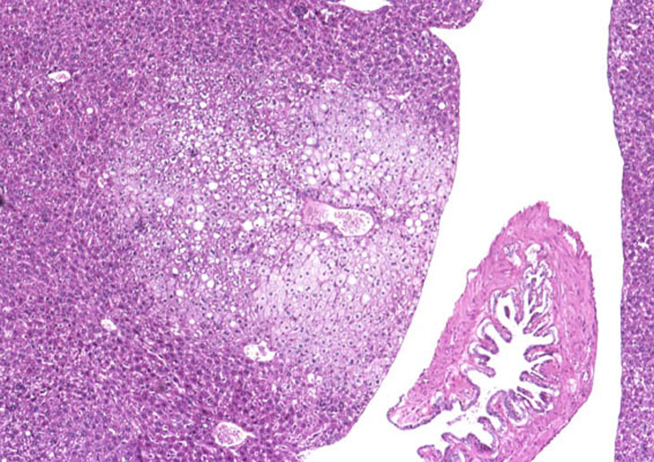
This clear cell focus actually has light pink staining of the cytoplasm.
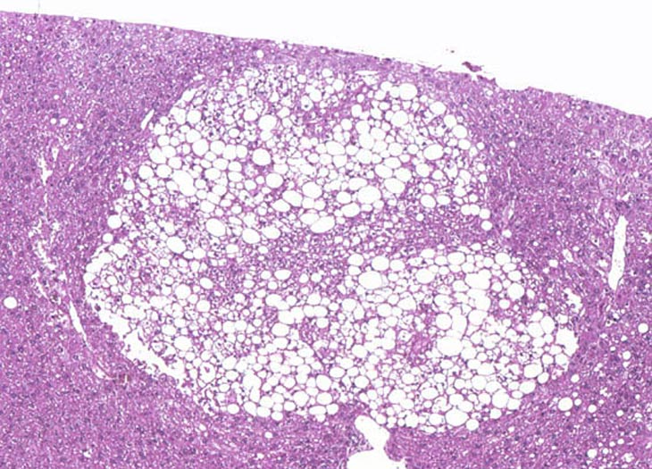
A vacuolated focus of cellular alteration comprised of a sharply demarcated collection of hepatocytes containing clear spaces. Some pathologists diagnosis this type of lesion as focal fatty change.
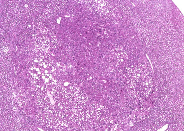
Mixed focus of cellular alteration. While there is a partial rim of basophilic hepatocytes, the central portion of this focus is comprised of a mixture of clear and amphophilic cells.
A large mixed focus of cellular alteration.
This focal lesion is believed to be associated with stress from the facliform ligament and resembles a focus of cellular alteration. These are examples of tension lipidosis and are not considered pathological changes.