


The Digitized Atlas of Mouse Liver Lesions
Much of the work carried out by DTT is in support of the National Toxicology Program (NTP), an interagency partnership of the Food and Drug Administration, National Institute for Occupational Safety and Health, and NIEHS.
Visit the NTP Website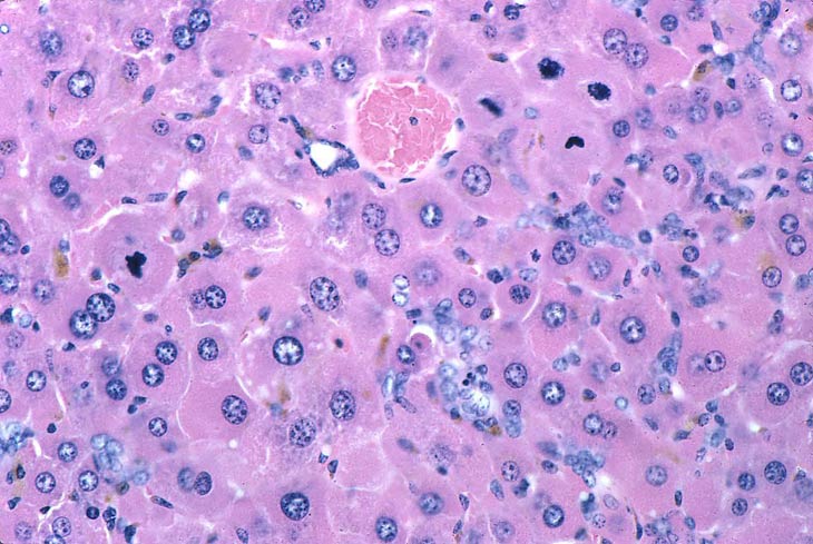
Large eosinophilic hepatocytes (eosinophilic granular cytoplasm represents peroxisome proliferation), biliary hyperplasia (ducts, ductules, oval cells), several mitotic figures, and pigment are present in the liver of this mouse fed a diet containing di-(2-ethylhexyl)-phthalate (DEHP) for 10 months.
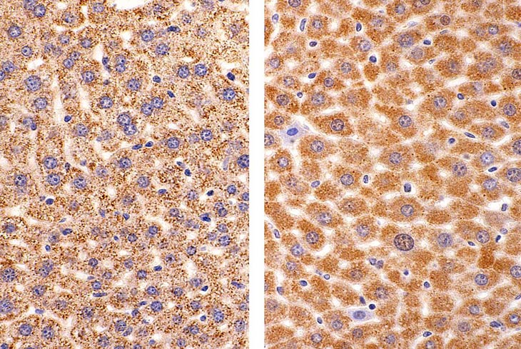
Oxidase immunohistochemical stain showing small oxidase-containing peroxisomes in smaller hepatocytes (on left) in a PPAR alpha null mouse given di(2-ethylhexyl)phthalate as compared with enlarged hepatocytes (on right) with peroxisome proliferation induced by DEHP in a wild type mouse.