


The Digitized Atlas of Mouse Liver Lesions
Much of the work carried out by DTT is in support of the National Toxicology Program (NTP), an interagency partnership of the Food and Drug Administration, National Institute for Occupational Safety and Health, and NIEHS.
Visit the NTP WebsiteApoptosis is a form of necrosis and is sometimes referred to as single cell necrosis. Apoptotic cells and apoptotic bodies in the liver are classically characterized by a rounding up of dying hepatocytes that often have hypereosinophilic cytoplasm and are surrounded by a clear halo resulting from fragmentation and contraction of the dying hepatocyte. Prior to development of rounded apoptotic cells and bodies, affected cells are hypereosinophilic, but may be more angular without a surrounding clear halo. Apoptotic bodies often contain fragments of dense nuclear material, which is hyperbasophilic, although some apoptotic bodies may consist of only condensed cytoplasm. Up to three adjacent apoptotic bodies are generally considered to originate from a single dying hepatocyte. Apoptotic bodies may be free or contained within Kupffer cells or normal hepatocytes.
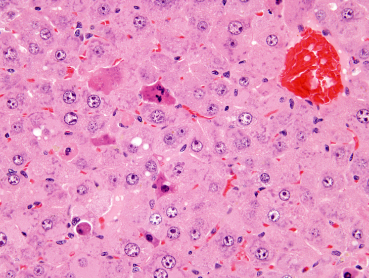
Early development of apoptosis. Affected hepatocytes are hypereosinophilic and angular with fragmented and condensed nuclear material within the cytoplasm.
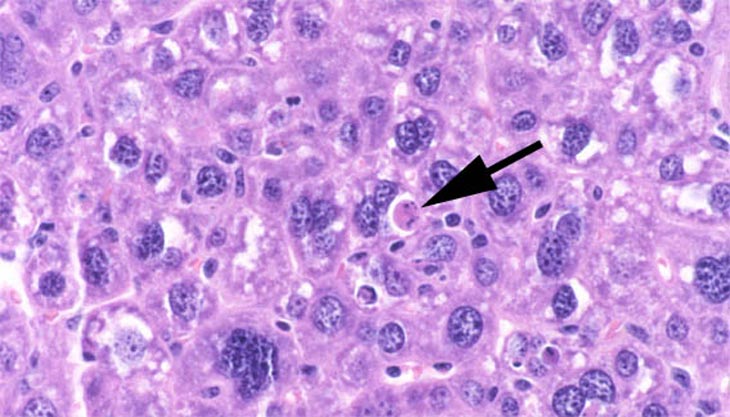
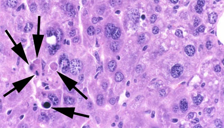
Apoptotic bodies (arrows) as well as polyploid hepatocytes are present. Polyploid hepatocytes containing either single large nuclei or multiple nuclei are also evident.
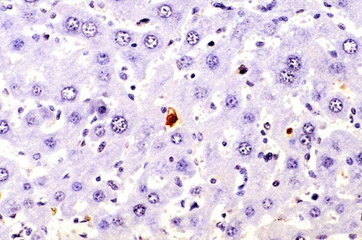
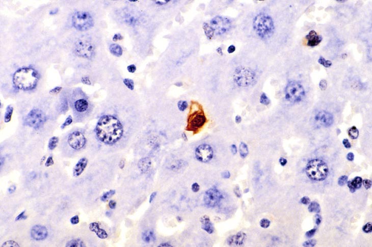
In situ end labeled of an apoptotic cell. Terminal desoxyribosyl-transferase mediated dUTP end labeling stain.