


The Digitized Atlas of Mouse Liver Lesions
Much of the work carried out by DTT is in support of the National Toxicology Program (NTP), an interagency partnership of the Food and Drug Administration, National Institute for Occupational Safety and Health, and NIEHS.
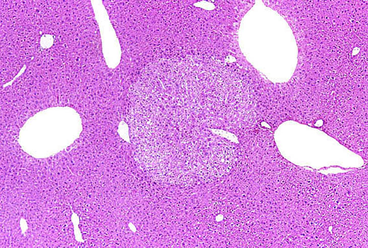
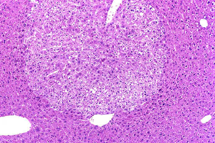
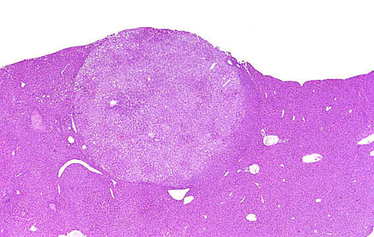
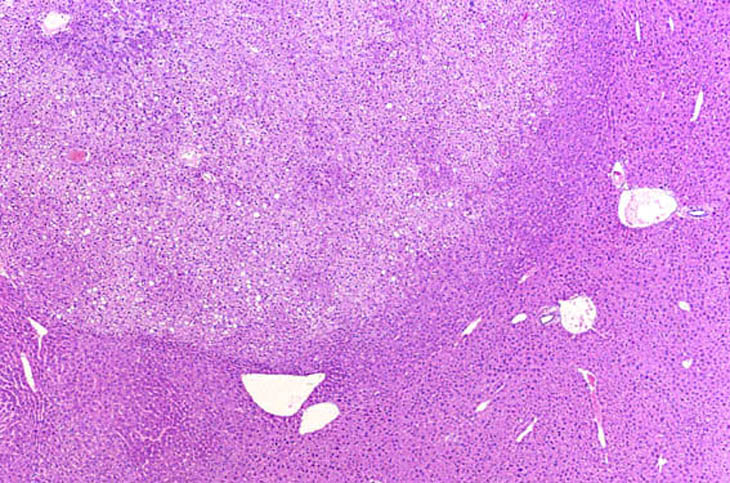
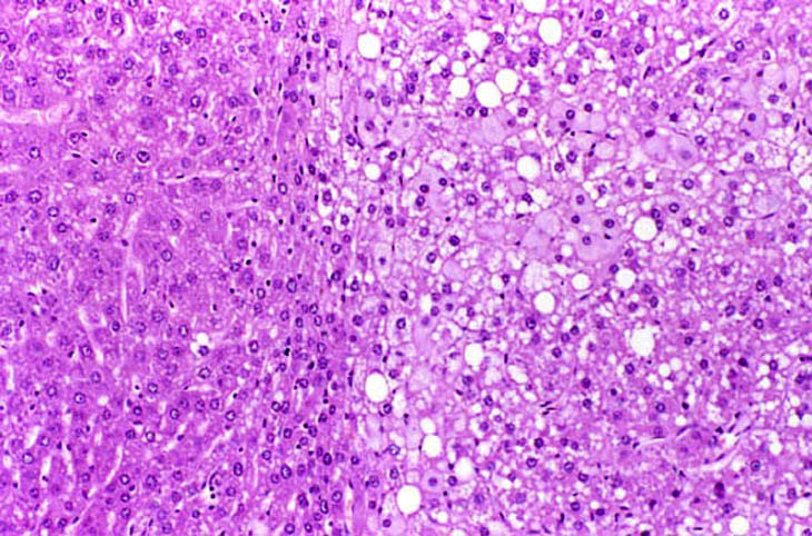
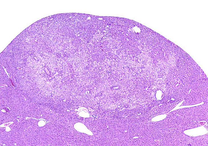
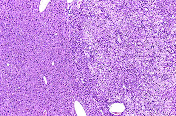
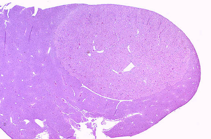
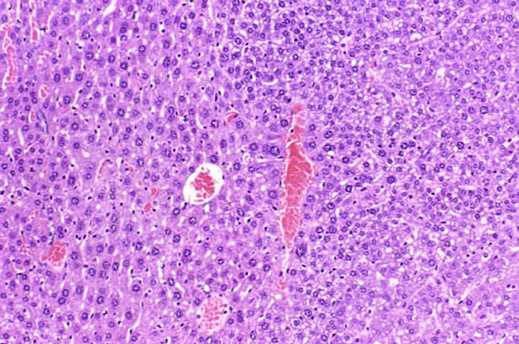
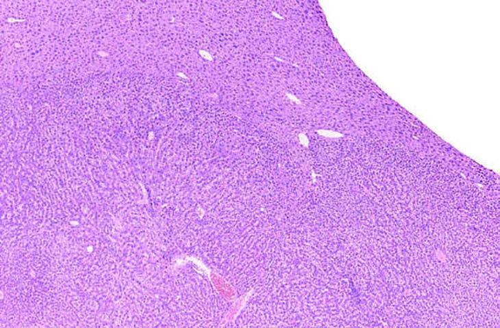
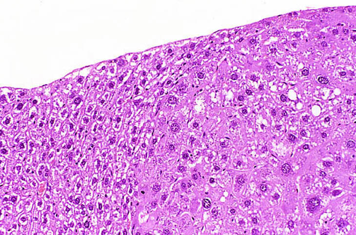
Visit the NTP WebsiteHepatocellular adenomas are typically discrete proliferative lesions with loss of normal lobular architecture, absence of portal areas, and some degree of compression of adjacent normal hepatic parenchyma. The arrangement of the hepatocytes within an adenoma may be at right angles to the surrounding hepatic parenchyma. The tinctorial properties of hepatocytes within an adenoma may be basophilic, eosinophilic, pale, or mixed.
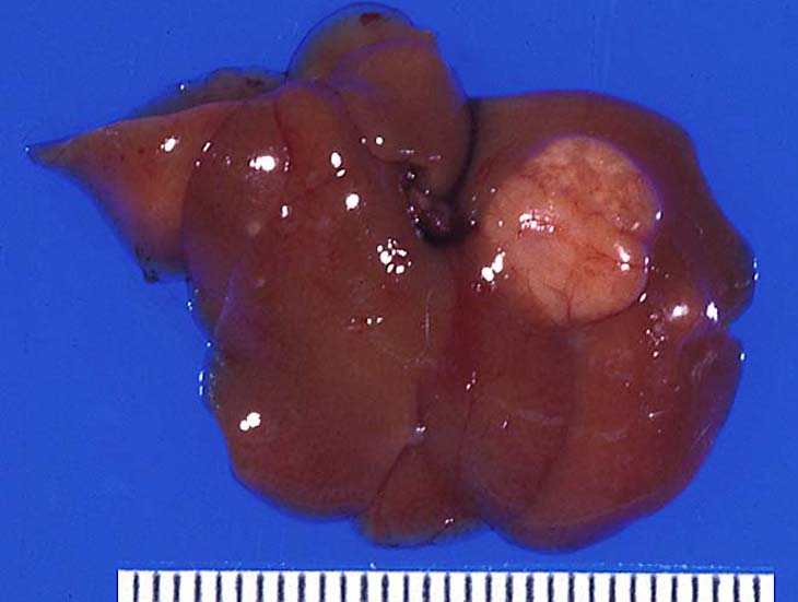
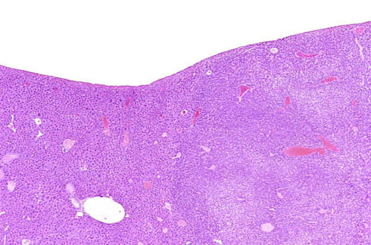
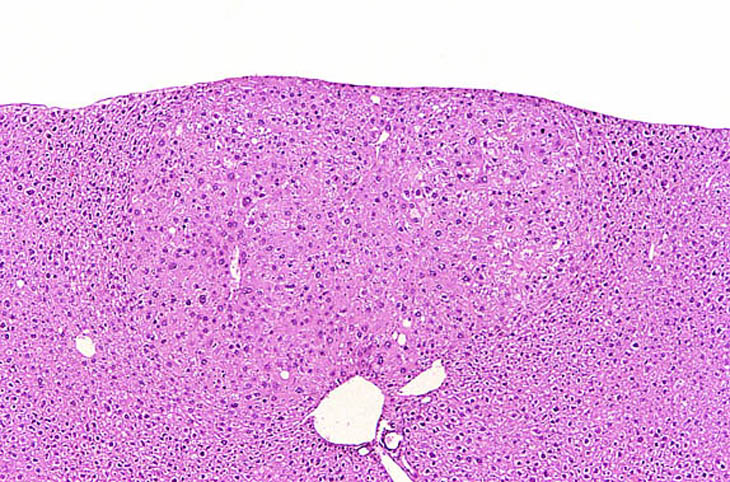
A large hepatocellular adenoma is present on the parietal surface of the left lobe.
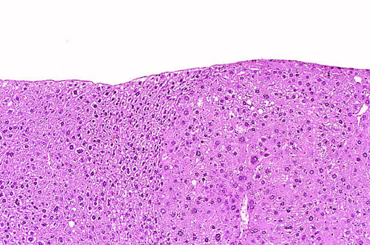
A discrete hepatocellular adenoma with pale staining cytoplasm.
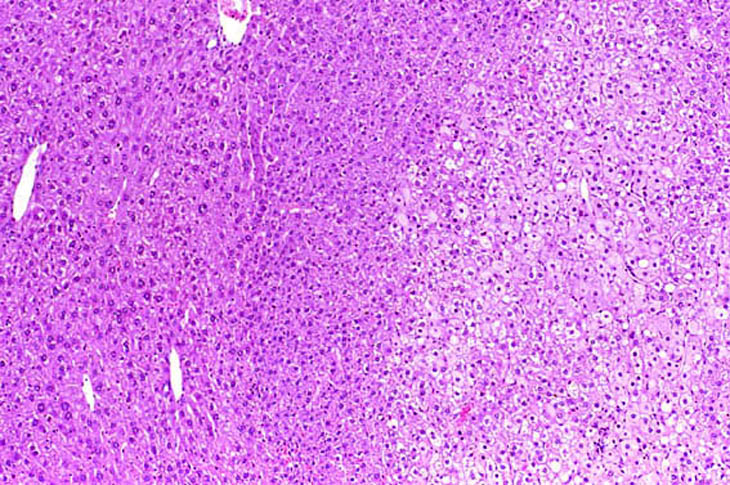
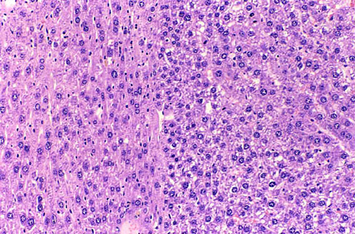
This adenoma has a partial rim of basophilic cells while the remaining cells are pale.
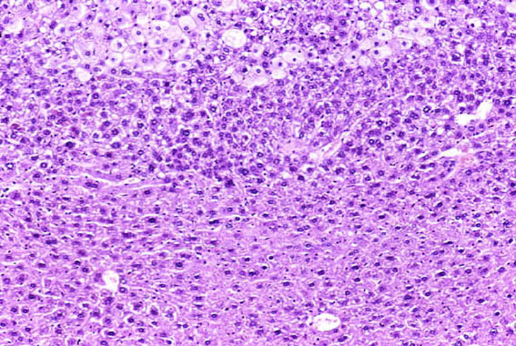
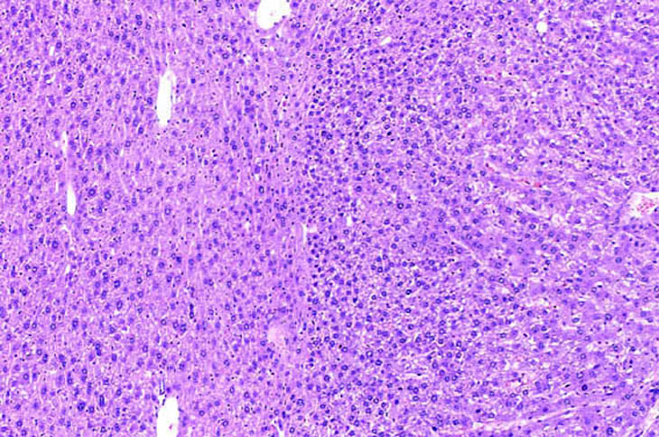
This mixed hepatocellular adenoma is comprised of an admixture of basophilic and pale staining hepatocytes.
A discrete eosinophilic adenoma.
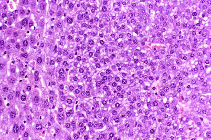
A basophilic adenoma. Basophilic adenomas are often comprised of closely spaced hepatocytes with small amounts of cytoplasm.
A basophilic adenoma.
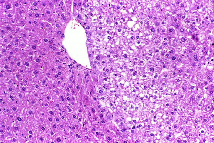
An eosinophilic adenoma. Note the abundant eosinophilic cytoplasm in contrast with the basophilic adenomas.