


The Digitized Atlas of Mouse Liver Lesions
Much of the work carried out by DTT is in support of the National Toxicology Program (NTP), an interagency partnership of the Food and Drug Administration, National Institute for Occupational Safety and Health, and NIEHS.
Visit the NTP WebsiteHemangiomas and hemangiosarcomas may arise as primary vascular neoplasms in the liver. They are usually not sharply demarcated from the surrounding parenchyma. Hemangiomas are recognized by a modest proliferation of a single layer of flatten, normal appearing endothelial cells lining vascular spaces with varying degrees of attendant hepatic cord atrophy. Hemangiosarcomas may arise as primary neoplasms in the liver, as well as in other tissues. The neoplastic cells are generally elongated or spindle-shaped and may form solid areas occupying dilated hepatic sinusoids. These neoplasms are typically locally invasive.
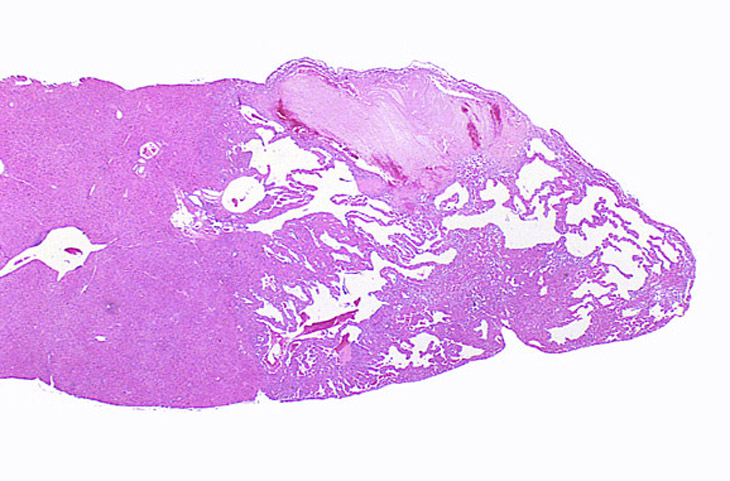

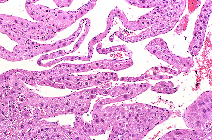
A hemangioma has destroyed the normal hepatic parenchyma. A thrombus is present at the top of the hemangioma; high magnification shows dilated vascular channels lined by flattened endothelial cells. There is minimal atrophy of hepatic cords.
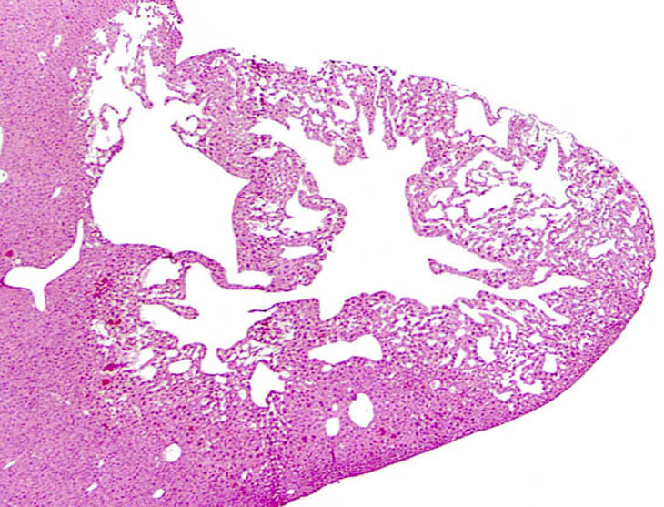
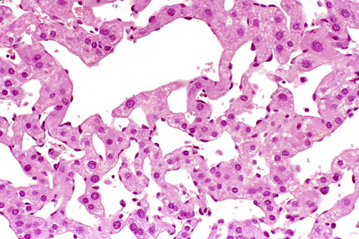
Dilated vascular spaces have replaced hepatic parenchyma in this hemangioma. High magnification shows dilated vascular spaces lined by flattened and sometimes dome-shaped endothelial cells and mild atrophy of the visible hepatic cords.
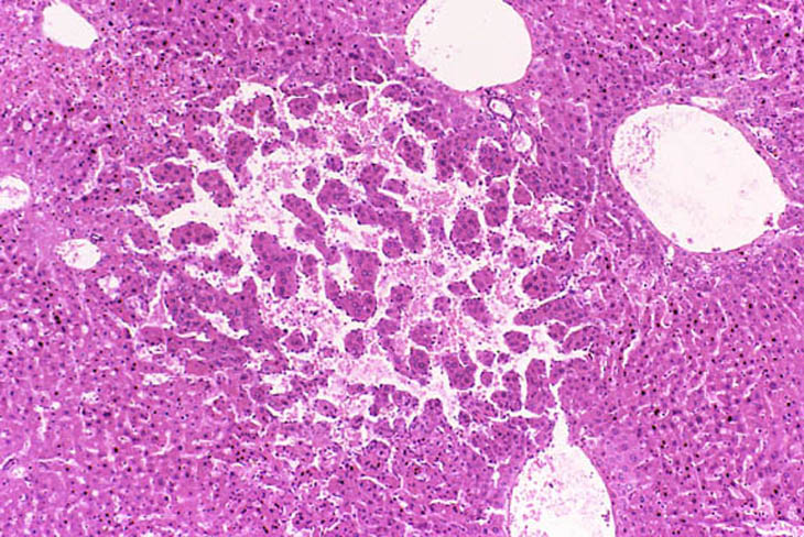
This hemangioma has isolated small clusters of hepatocytes; higher magnification shows proliferation of flattened endothelial cells.
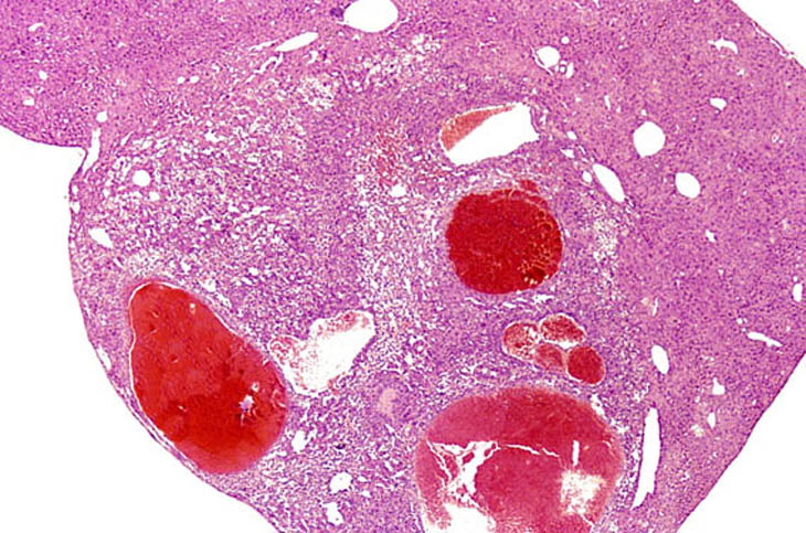
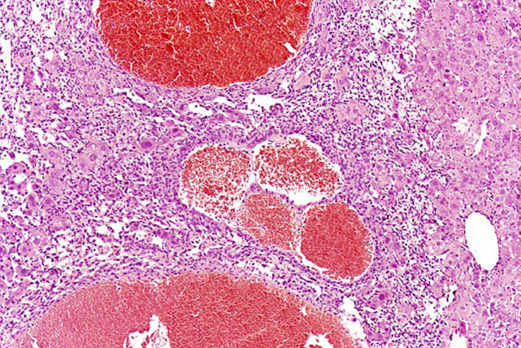
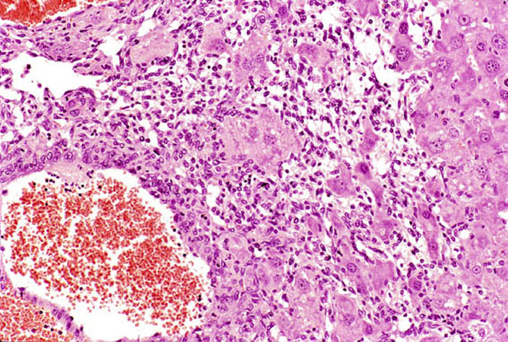
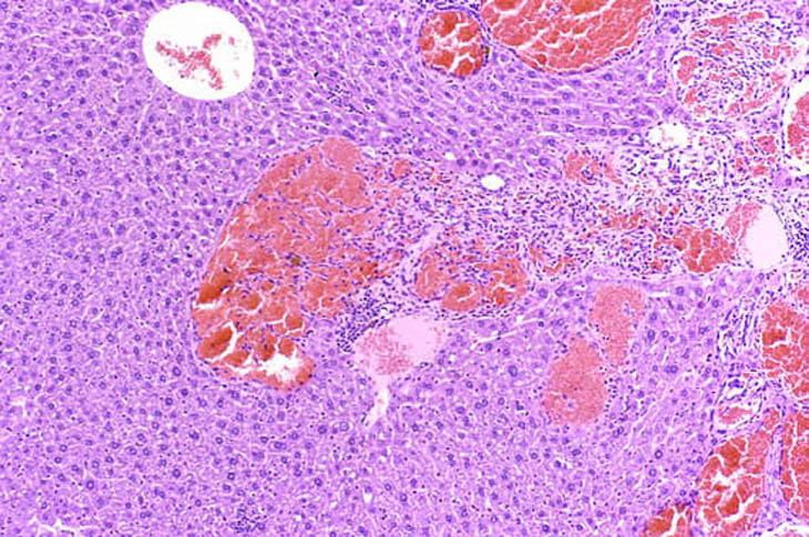
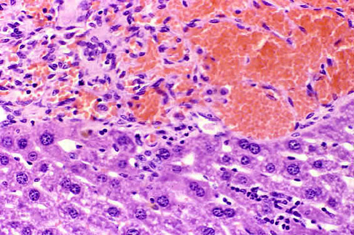
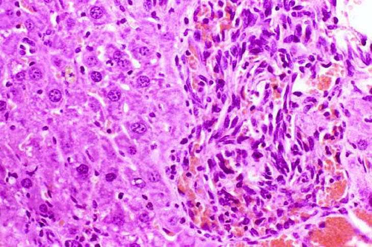
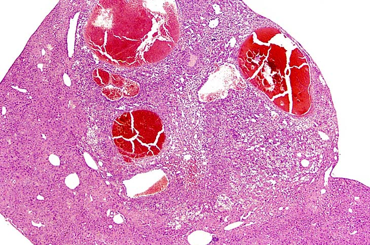
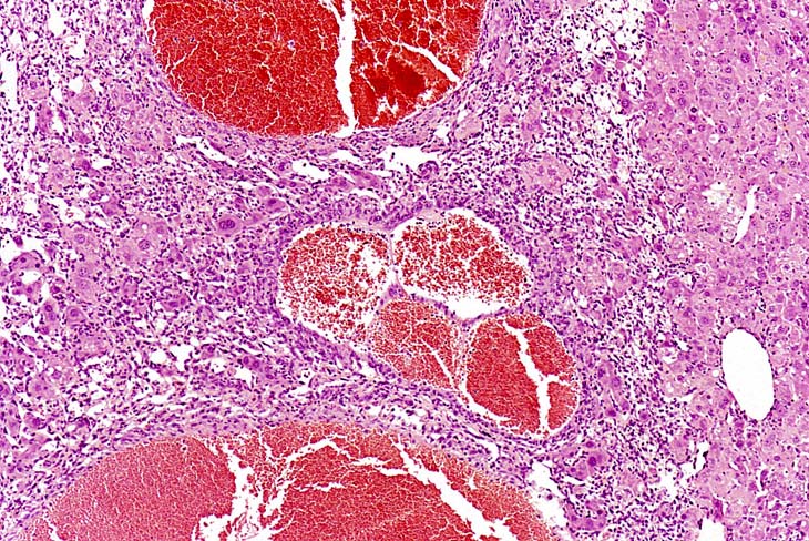
This hemangiosarcoma is well demarcated from the normal hepatic parenchyma; sharp demarcation is an unusual feature of hemangiosarcomas. Large blood-filled lakes are apparent. Higher magnification shows bands of spindle-shaped endothelial cells with attendant destruction of hepatocytes, proliferation of plump endothelial cells, and atrophy of hepatocytes.
Another example of a hemangiosarcoma.
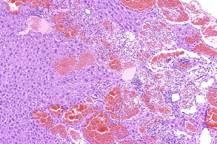
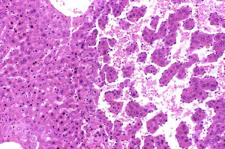
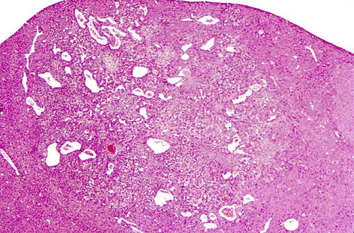
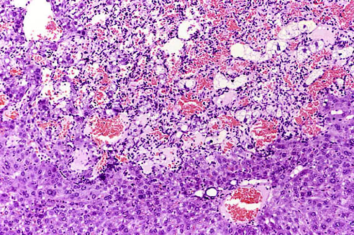
This hemangiosarcoma is poorly demarcated from surrounding parenchyma and consists of dilated blood-filled spaces and sheets of proliferating endothelial cells.
Hemangiosarcoma in an adult mouse.
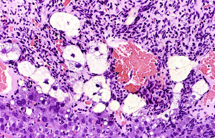
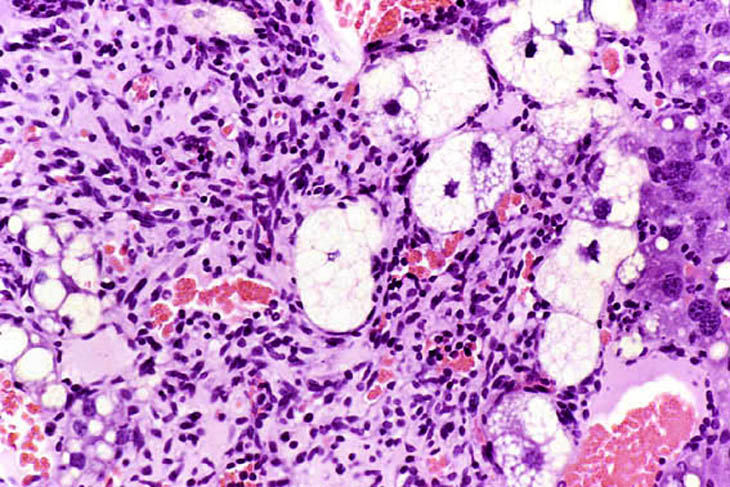
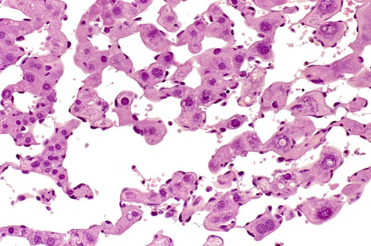
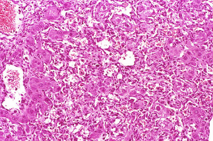
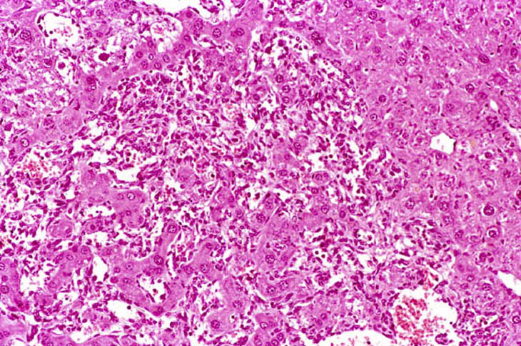
This hemangiosarcoma is has caused degenerative changes in some of the entrapped hepatocytes. The degenerating hepatocytes are enlarged and vacuolated with evidence of nuclear degeneration.
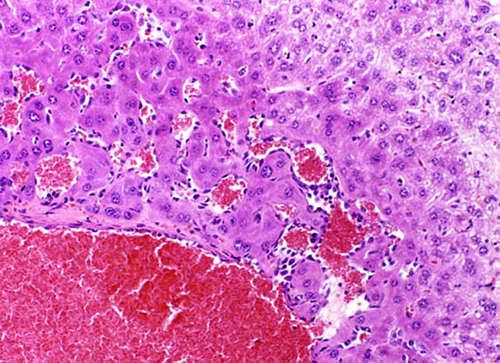
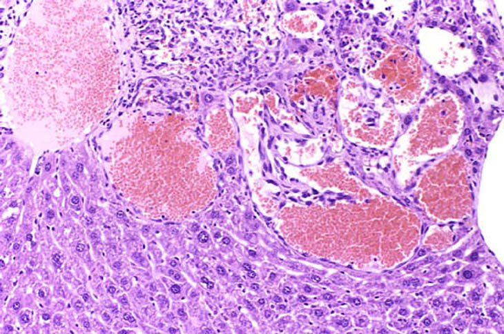
This lesion appears consistent with early endothelial cell proliferation and peliosis but is actually at the edge of an hemangiosarcoma.

Another example of an hemangiosarcoma from a male B6C3F1 mouse.